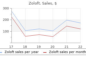
Zoloft dosages: 100 mg, 50 mg, 25 mg
Zoloft packs: 30 pills, 60 pills, 90 pills, 120 pills, 180 pills, 270 pills, 360 pills

Order cheap zoloft on lineIn all circumstances depression definition nih purchase zoloft with amex, the design of a contrast agent have to be accompanied by the design of a suitable management agent to allow assessment of the specificity of the compound anxiety attacks symptoms treatment buy zoloft 100 mg visa. Ideally neonatal depression definition cheap 50 mg zoloft with mastercard, the management agent have to be as much like depression definition duration generic zoloft 100mg free shipping the distinction agent as attainable to avoid confounds linked to broadly totally different pharmacokinetic properties, diffusion properties, and so forth. Genetic modifications to cancer cells that make them overexpress ferritin receptors are particularly appealing to the examine of most cancers metastasis. In fact, points associated to label dilution or to delivery of exogenous distinction brokers to particular cells are principally alleviated since these cells are in a place to generate their very own iron monocrystals from out there iron in their microenvironment. Macromolecular therapeutics-Advantages and prospects with special emphasis on strong tumour focusing on. Blood circulate, metabolism, mobile surroundings, and progress rate of human tumor xenografts. The design of a distinction agent is dictated by several crucial factors, corresponding to the location of the target. Metabolic trapping as a principle of radiopharmaceutical design: Some elements responsible for the biodistribution of [18F] 2-deoxy-2-fluoro-d-glucose. Brain tumor enhancement in magnetic resonance imaging dependency on the extent of protein binding of applied contrast agents. Classification and basic properties of contrast agents for magnetic resonance imaging. A "good' magnetic resonance imaging agent that reports on particular enzymatic activity. New enzyme-activated solubility-switchable contrast agent for magnetic resonance imaging: From synthesis to in vivo imaging. Paramagnetic polymerized liposomes: Synthesis, characterization, and functions for magnetic resonance imaging. Detection of tumor angiogenesis in vivo by v3-targeted magnetic resonance imaging. Magnetic resonance distinction enhancement of neovasculature with alpha(v)beta(3)-targeted nanoparticles. Molecular imaging of angiogenesis in nascent vx-2 rabbit tumors using a novel v3-targeted nanoparticle and 1. Highefficiency intracellular magnetic labeling with novel superparamagnetic-Tat peptide conjugates. Specific focusing on of breast tumor by octreotide-conjugated ultrasmall superparamagnetic iron oxide particles using a scientific three. In vivo offresonance saturation magnetic resonance imaging of v3targeted superparamagnetic nanoparticles. Molecular imaging of pancreatic cancer in an animal model using focused multifunctional nanoparticles. Magnetic nanoparticles for early detection of most cancers by magnetic resonance imaging. Bridot J-L, Faure A-C, Laurent S, Riviere C, Billotey C, Hiba B, Janier M, Josserand V, Coll J-L, Vander Elst L, Muller R, Roux S, Perriat P, Tillement O. Hybrid gadolinium oxide nanoparticles: Multimodal contrast agents for in vivo imaging. Chemical change saturation switch contrast agents for magnetic resonance imaging. Chauvin T, Durand P, Bernier M, Meudal H, Doan B-T, Noury F, Badet B, Beloeil J-C, T�th �. Synthesis of gadolinium-labeled shell-crosslinked nanoparticles for magnetic resonance imaging purposes. In vivo characterization of activatable cell penetrating peptides for targeting protease exercise in cancer. Splenic imaging with 99mTc-labeled erythrocytes: A comparative study of cell-damaging methods. Human lymphocyte site visitors assessed by indium-111 oxine labelling: Clinical observations. Application of the static dephasing regime concept to superparamagnetic iron-oxide loaded cells. Positive distinction magnetic resonance imaging of cells labeled with magnetic nanoparticles. Imaging adenoviral-directed reporter gene expression in dwelling animals with positron emission tomography. For example, a normal estimate from the thermal Boltzmann population exhibits that detectable spins can be increased from about 1 to 28 elements per million (scaled linearly) when going from zero. While present fields are adequate for acquiring info from protons within the millimolar focus range. These advances have enabled the penetration of latest layers of prognosis and have driven the emergence of new functions. This research provides clear evidence that hyperpolarized pyruvate and its metabolites are preserved lengthy sufficient in vivo to be imaged. It additionally demonstrates that differential conversion of pyruvate between tumor and normal tissue can be imaged from a normal carotid injection. In summary, the brokers only last a couple of minutes in favorable circumstances, but helpful information from tumor metabolism could be obtained in as little as 10 s. In addition to being technically tough, this causes a major discount in polarization because of T1 leisure throughout warming. In addition, current work [12,13] has demonstrated the feasibility of preparing and imaging hyperpolarized Suc and Gln in vivo and in vitro, respectively. Microwave radiation is then used to saturate the electron spin states of the radical in the solid state, facilitating the transfer of polarization from electrons to target nuclei. Frozen hyperpolarized target compounds are then launched into the answer after rapid (~1 s) dissolution. Rapid dissolution is important to preserving nuclear spin polarization and making the hyperpolarized tracers injectable for in vivo use. A typical process for preparing hyperpolarized pyruvate by these methods consists of mixing an answer containing the target metabolic agent with a trityl radical/glycerol matrix that permits for efficient polarization switch from radical free electron to target nucleus corresponding to 13C or 15N. The pattern is then quickly thawed by mixing with scorching water, and the ensuing answer is then obtainable for injection into an organism. Parahydrogen may be routinely produced by passing high-purity hydrogen (25% para and 75% ortho at room temperature) gasoline through a catalyst-filled column at cryogenic temperatures in the vary of 5�20 K, permitting quick conversion of the ortho-/para- mixture of hydrogen fuel to parahydrogen. Production of parahydrogen fuel can subsequently be scaled up significantly to accommodate foreseeable scientific demands [24]. In addition to simple preparation, parahydrogen undergoes back-conversion to orthohydrogen very slowly, on the time scale of weeks, and can be stored for durations of weeks to months at room temperature. As of 2010, this hyperpolarization technology is pursued in basic and translational analysis by a number of teams [5,13,28�30]. As a end result, polarization losses associated with the spin lattice relaxation time (T1) of X nuclei and the decay of the singlet state of the hooked up spins are minimized. Moreover, the water-soluble catalysts enable the complete hyperpolarization procedure to be performed in an aqueous medium, a requirement for in vivo purposes. This molecular addition requires a catalyst and often is carried out on the time scale of some seconds by injection of an aqueous answer containing molecular precursors and the catalyst within the ambiance of parahydrogen gasoline (1�12 atm). The efficiency of the polarization switch [32] is dependent upon scalar couplings of the concerned nuclei and requires prior data of the spin�spin couplings between these three nuclei; the related couplings may be decided at physiological pH and used with out further refinement. Moreover, the molecular tracer is typically deuterated to have the ability to simplify the spin system and to increase rest occasions. While hyperpolarized 129Xe has found utility in lung and mind imaging, it has not yet been effectively utilized as a potential polarization reservoir for metabolic cancer applications as a outcome of the difficulty of efficient polarization transfer to 13C and 15N [37]. This inherent issue arises as a outcome of the transfer mechanism requires extended close contact between the isotopes. This has motivated the scientific neighborhood to immobilize hyperpolarized xenon in cages in proximity to modified moieties for functionalization [38], as commonly performed in molecular imaging such as gadolinium-based chelates [39]. In distinction, the choice approach is to switch the spin polarization of 129Xe to unhazardous metabolites previous to injection. Mechanisms for polarization switch that exploit relatively weak spin�spin (usually referred to as J-) couplings are usually excluded, as a outcome of covalent linkages with 129Xe are precluded because of the inert nature of xenon.
Purchase genuine zoloft onlineInternational Journal of Radiation Oncology bipolar depression symptoms test free order genuine zoloft on-line, Biology mood disorders symposium johns hopkins buy generic zoloft 25mg line, Physics 2001;49(4):1097�1108 depression symptoms pressure head buy generic zoloft 50mg line. Current issues within the utility of 19F nuclear magnetic resonance methodologies for the assessment of tumour hypoxia depression prevention order zoloft cheap. Quantitative assessment of blood flow, blood volume and blood oxygenation effects in practical magnetic resonance imaging. Magnetic Resonance Materials in Physics Biology and Medicine 2004;17(3�6):288�295. Effects of respiratory a hyperoxic hypercapnic gas mixture on blood oxygenation and vascularity of head-and-neck tumors as measured by magnetic resonance imaging. International Journal of Radiation Oncology, Biology, Physics 2002;53(5):1185�1191. International Journal of Radiation Oncology, Biology, Physics 2007;68(4):1065�1071. In on a daily basis medical follow, tumor response evaluation is a relatively informal and qualitative process. Imaging research are performed at baseline some time prior to the start of therapy, after which again at interval follow-up periods some (biologically appropriate) time after the beginning of treatment. Follow-up research are typically performed every 6�12 weeks, relying upon the pattern of remedy cycles. The radiologist reporting the follow-up case usually compares the imaging study to the most recent examine and reports qualitative modifications in overall tumor burden as typically increasing, lowering, or remaining stable. The goal of therapy response assessment is to categorize the efficacy or toxicity of a treatment for an individual affected person or affected person cohort. Serial imaging studies are used to assess adjustments in the location, dimension, and metabolic activity of tumors over time. In the superior illness setting, tumors typically shrink or stay secure in dimension with remedy for some time frame however ultimately acquire mechanisms of resistance that allow them to develop once more. The oncologist uses the images, radiology stories, and additional medical features, such as patient toxicity to therapy, to decide if treatment should be continued or discontinued. If the tumor burden is increasing or there are new lesions current, then the treatment is discontinued and new treatment options are thought of. On the opposite hand, for sufferers taking part in therapeutic medical trials, the response evaluation course of is much more formalized and quantitative. Novel most cancers therapies are often evaluated first within the metastatic or advanced disease setting for his or her antitumor activity. The major therapeutic goal for many antitumor remedies within the superior disease setting is delay in tumor development and ideally tumor shrinkage. Delay in tumor progress can correlate with improved high quality of life, morbidity, and mortality [1,2]. Historically, tumor shrinkage has been the hallmark of antitumor activity for cytotoxic therapies, which trigger tumor cell death, and thus have the potential to shrink tumor plenty. Noncytotoxic therapies on the other hand are sometimes cytostatic, and may not cause tumor shrinkage however rather tumor stability. Several noncytotoxic therapies have additionally demonstrated improvement in total survival in randomized trials [5,6]. For such cases, delay in tumor growth can be used as evidence of antitumor activity [7]. The aim of therapeutic scientific trials is, thus, to decide if an experimental therapy is efficacious and safe. In order to evaluate the efficacy and toxicity of the experimental treatment among the many sufferers within a medical trial, the trial protocol is developed to standardize treatment and response evaluation procedures for all taking part topics. Formal response standards have been developed to assist standardize tumor response evaluation across medical trials so that trial results may be in contrast. For each medical trial, the response evaluation protocol particularly defines the imaging modality, image acquisition protocol, the timing of the baseline and follow-up assessments, and the response criteria that should be used to quantify and classify response. Cancer response standards standardize the approach for estimating tumor burden, defining quantitative and qualitative adjustments in tumor burden, and classifying tumor response to treatment in clinical trial cohorts. These formal response evaluation outcomes are incorporated into the scientific trial determination algorithms that define the situations when an experimental remedy must be continued or discontinued. In this fashion, the scientific trial protocol helps to guarantee consistent treatment decisions across trial subjects. Response charges beneath zero on the y-axis correspond to partial tumor shrinkage with therapy, while these above zero correspond to tumor development. The finest percent change in the tumor burden from baseline (y-axis) is plotted for each patient in the cohort (x-axis). The mean of the quantitative response fee may additionally be taken and in contrast between clinical trial arms as a quantitative estimate of variations in remedy efficacy. Trial arms may also examine the median time to progression and the percentage of topics with a selected response class. A clinically and statistically important distinction within the time to development between two trial arms is usually used as an intermediate endpoint for novel drug approval by regulatory businesses. This enables more rapid approval of novel therapies than waiting for overall survival endpoints to be reached, which for many cancers may be a number of years. While scientific signs of the illness are an necessary aspect of assessing particular person affected person responses, they have an inclination to be subjective assessments and never all patients have such symptoms. The quantitative response was calculated as the percent change in the tumor burden (sum of products), taking as reference the baseline evaluation. Response Rate = (Tumor Burden follow-up - Tumor Burden baseline) Tumor Burden baseline and compare trial outcomes [9,10]. In the mid-1990s a global, multidisciplinary committee known as the International Working Party was established to simplify and standardize the standards. First, it defined more specifically what was thought-about measurable illness each by the anatomic location of the lesion and by a minimal size requirement. Second, as a lot as five lesions per organ could possibly be chosen with a maximum of 10 lesions complete to estimate tumor burden. Lastly, the factors utilized unidimensional measurement of lesions and the sum of the longest diameters to estimate total anatomic tumor burden. As a results of changing from bidimensional to unidimensional measurement, the thresholds for outlining response classes were also modified. Major changes include particular definitions for what is taken into account measurable with respect to lymph nodes, and a lower within the variety of target lesions used to estimate tumor burden from 10 total to five whole with a maximum of two lesions per organ. Two generations of response the quantitative response was calculated at each follow-up evaluation period and the minimum tumor burden achieved, since baseline was used to calculate the best response rate. The response classes outlined and in use to this present day are complete response, partial response, stable disease, and disease development. With qualitative descriptions, these response categories are forms of ordinal scale. The criteria also specified observations together with the appearance of any new lesions as defining the event of illness progression. However, the designers of medical trials started to make modifications to the standards on an advert hoc foundation, to incorporate new imaging applied sciences and address underspecified aspects of the original document. As such, response criteria have been developed for these specific illnesses [17,21]. Once the affected person is enrolled in the trial and previous to starting therapy, a baseline imaging examine is required to assess the areas and sizes of the tumor lesions. In the current scientific workflow, the oncologist orders an imaging study, the patient has the examine carried out and the radiologist is the primary to evaluation the pictures. The radiologist summarizes her/his findings in a textual content report and records detailed measurements as image markups. The green markings are an instance of the image markups created by the radiologist that measures the longest diameter and its respective perpendicular (short) axis. The report is then despatched to the oncologist who independently critiques the report and pictures. Tumor burden is assessed both quantitatively and qualitatively, depending upon the location of the tumor lesion and the kind of imaging modality. Quantitative measurements are recorded where applicable; in any other case, a Boolean description is made, denoting the persistence or resolution of a lesion.
Diseases - Hyperornithinemia, hyperammonemia, homocitrullinuria syndrome
- Emery Dreifuss muscular dystrophy, dominant type
- Esophageal disorder
- Dystonia musculorum deformans
- Microcephalic primordial dwarfism Toriello type
- Glucagonoma
- Human ewingii ehrlichiosis
- Pallister Hall syndrome
100mg zoloftThey quantified the accuracy of their segmentation results by way of comparison to manual segmentation by an expert bipolar depression for a year hoping for mania buy 100mg zoloft with mastercard. Recall that atlas-based approaches rely on the technology of a 2D or 3D atlas that represents the "typical" topic mood disorder teenager buy zoloft 25mg visa. This atlas is developed by imaging a inhabitants of "regular" topics and then merging the ensuing image data sets into a single image depression urban dictionary purchase zoloft 100 mg with amex, or atlas anxiety and pregnancy purchase generic zoloft online, that best represents the inhabitants. Target patient scans had been then registered to the atlas utilizing a mixture of affine and nonlinear registration methods. The brainstem, parotids, and mandible had been then segmented so that the size and place of those constructions might be measured to decide an effective plan for radiation remedy that would avoid radiation exposure of important organs. Even if these strategies are used, they normally indicate the places of "suspicious" regions, and the ultimate decision is made by the physician. Breast cancer screening has been a fertile area for automatic segmentation analysis for cancer detection, and several other examples of laptop aided prognosis are presented later on this section. The following text is organized by cancer sort, and examples of developments are introduced for most cancers of the breast, prostate, and mind. There are a wide range of causes for this, however probably the most distinguished motivation is that based on the American Cancer Society, 1 out of each 8 ladies (12. This study included 19 ladies and 39 marked lesions, that are used as floor truth to consider algorithm efficiency. As ordinary, nevertheless, tuning an algorithm to be this sensitive comes with a corresponding worth of a comparatively low specificity. The authors report a 10% false detection fee per slice, which is problematic in that it can probably result in a large quantity of pointless biopsies. The authors report that their outcomes compared with human segmentation and reported a tumor segmentation confidence of 95%. Prostate cancer can be a popular target for computer-aided analysis due to the precise fact published by the American Cancer Society that about 1 in each 6 men shall be recognized with prostate most cancers in their lifetime (see. A few particular and up to date examples of this might be presented here, however a more thorough historical evaluation of computer-aided diagnostic instruments for prostate most cancers, including automated segmentation technologies, may be present in Zhu, Williams, and Zwiggelaar (2006). Their outcomes showed equal or higher performance on most cancers detection as in comparison with experts on a case examine that included 33 patients. Because of the excessive sensitivity of multiple organs in the head/neck area, exact location and quantification of tumor volume is required for nasopharyngeal instances throughout treatment. One requirement of a region growing strategy is the position of a seed point someplace in the tumor, and that is usually carried out manually by an skilled. The requirement to place a seed point within the tumor renders area rising an ineffective segmentation strategy for cancer detection. A vary of 86% to 94% match was reported by comparing the automated tumor segmentation outcomes to handbook outcomes captured by an expert radiologist. There has been a big investigative effort of segmentation methods for mind cancer staging. It is predicated on a multiscale Bayesian strategy to perform tissue classification (tumor versus nontumor). The paper acknowledges that although a lot progress has been made within the last decade on automated tools for tumor staging, only a few are clinic ready. An algorithm for mind tumor segmentation is presented in Wang, Cheng, and Basu (2009) that stories a model new lively contour method that improves seize range. The paper presents a examine using picture knowledge from the Internet mind segmentation repository Segmentation strategies based mostly on level units are also commonly used for brain tumors. To overcome some of the challenges with setting up the pace perform, a way that includes a dynamically up to date pace perform is presented in Taheri, Ong, and Chong (2009). Typically, level sets depend on boundary information for convergence, but an approach that includes each region and boundary information concurrently. An illustrative example of an algorithm that presents a strategy for making use of a quantity of scans with various spatial resolution is offered in Nie et al. The algorithm was utilized to malignant glioma mind tumor segmentation and outcomes were similar to manually segmented tumor results by a quantity of subject matter consultants. First, handbook segmentation is susceptible to inconsistency, even when carried out by specialists. The second and more vital problem is that correct "floor reality" data are sometimes very onerous to come by for many particular functions of curiosity. However, by choosing algorithms appropriate to a specific problem, one can present environment friendly complementary instruments to assist clinicians in cumbersome actions such as finding and delineating the tissue regions present in the images. In the next, a big selection of examples are offered to illustrate how region-, model-, or contour-based picture processing algorithms can contribute in cancer treatment planning. One must (1) acquire the cranial and mind volume measurements, (2) localize gray and white and encephalic liquid as nicely, and (3) determine and localize potential lesions. Usually performed utilizing commonplace parametric strategies and Gaussian mixture models, the writer identifies a limitation and lack of efficiency of these approaches within the case of an rising variety of elements together with presence of abnormal tissues. Moving towards a nonparametric model, the writer presents a Bayesian strategy primarily based on the Dirichlet combination mannequin. An essential function of this mannequin is that inference on the number of parts within the mixture is incorporated, a Markov random area offers some noise immunity as nicely. As expected, these strategies carried out nicely in segmenting irregular tissues characterized by significantly completely different texture traits in comparability with the environment. However, the segmentation accuracy was only reported to be 75% at finest (as compared to expert handbook results), so it appears troublesome to delineate precisely the entire tumor with these methods. The authors examine the performance/time ratio of those automated strategies against manual delineation. They note performance outcomes by method of a quantity overlap (a) (b) measurement, Vo, which is defined because the ratio of the intersection to the union of the automatic and manual segmentation quantity outcomes (optimal worth is 1. Simultaneous segmentation of each cortical surfaces is performed utilizing a coupled level-set strategy. For instance, for the automatic segmentation of the rectum on 44 total patients, 54% of the patient results have been thought-about to be "acceptable," "good," or "wonderful," while 45% were thought-about "not acceptable. Their method is designed to be flexible so that segmentation results (details of that are left to the paper) can be easily refined by tweaking the input parameters. Quantitative evaluation of the delineated space primarily based on the Jaccard similarity metric (Js) (Jaccard 1901) offers info of the therapy progress. At first, the picture is thresholded to extract the position of the liver, and then morphological operators are applied to extract the tumor candidates. Solomon, Butman, and Sood (2006) current the mix of a spatiotemporal mannequin and a Markov mannequin to perform fourdimensional mind tumor segmentation. Segmentation strategies range from data-driven (bottom-up) methods to model-driven (top-down) methods, and in addition embrace a number of strategies which would possibly be combos of both (hybrid techniques). For cancer-specific applications, the most generally researched and employed techniques embrace various types of pixel-based classification. Other approaches gaining recognition for brain segmentation are model-based methods corresponding to atlas-based segmentation. There is a wealth of research being printed in every of these categories, and the purpose of this chapter was to introduce the reader to probably the most promising strategies to permit them to (1) select a kind of segmentation strategy most applicable to their most cancers imaging analysis and (2) use the data as a beginning point for implementing their own segmentation techniques that construct upon the wealth of research being carried out. Multi-atlas based mostly segmentation of brain images: Atlas selection and its impact on accuracy. Magnetic resonance imaging follow-up of liver growth of neuroendocrine tumors in an experimental mouse model. Improving segmentation accuracy for magnetic resonance imaging utilizing a boosted choice tree. Automatic segmentation of magnetic resonance pictures utilizing a choice tree with spatial data. Volumetric texture analysis of breast lesions on contrastenhanced magnetic resonance photographs. Atlas-based segmentation of 3D cerebral constructions with competitive stage units and fuzzy management. Magnetic resonance imaging primarily based quantity estimation of ovarian tumours: Use of a segmentation and 3D reformation software program. Development of a unified probabilistic framework for segmentation and recognition of semi-rigid objects in complex backgrounds through deformable form fashions. A qualitative and a quantitative analysis of an auto-segmentation module for prostate cancer. �tude Comparative De La Distribution Florale Dans Une Portion Des Alpes Et Des Jura (Comparative study of the floral distribution in a portion of the Alpes and Jura regions). Segmentation of magnetic resonance images utilizing fuzzy algorithms from learning vector quantization.
Purchase zoloft 50 mg fast deliveryOver 90% of ankylosed teeth are deciduous anxiety guidelines generic zoloft 50mg with mastercard, most frequently depression knee pain cheap zoloft 100 mg overnight delivery, the second molar adopted by the primary molar depression symptoms ocd order zoloft now. Early ankylosis ends in noneruption or partial eruption depression test dass cheap zoloft 25 mg with mastercard, leading to incomplete growth of the alveolar course of. The failure of an ankylosed tooth to erupt may permit adjacent tooth to drift and allow supereruption of the tooth within the opposing arch. Panorex reveals incomplete eruption of major tooth with no permanent successor, indicating ankylosis. Periodontal standing: this is very important, because preexisting periodontal pathologies could be exacerbated throughout orthodontic and orthognathic surgical therapies. B, If tied into an lively straight arch wire, the adjoining tooth will be orthodontically moved toward the ankylosed tooth, resulting in the growth of a major malocclusion (in this case, an open chunk due to incisor intrusion). A lack of attached gingiva within the decrease incisor region before initiation of orthodontics if left untreated will cause severe gingival retraction and loss of supporting bone. Macroglossia could trigger anterior open chew, diastema between the teeth, accentuated maxillary curve of Spee, and reverse mandibular curve of Spee. Any pretreatment of acute or continual periodontal illness should be addressed earlier than the orthodontics and surgery. Gingival grafting could also be indicated before orthodontics to present hooked up gingiva in order to prevent these issues. Orthodontics can help put together interdental osteotomy websites by tipping the roots of the adjoining teeth away from each other to increase the interosseous area between the roots. The failure to recognize preexisting periodontal pathology, determine threat elements, poor performance of surgery, and/or lack of attention to detail may result in vital periodontal problems as well as different issues that might compromise the ultimate result. Tongue assessment: An enlarged tongue (macroglossia) can cause dentoskeletal deformities and instability of orthodontic and orthognathic surgical therapies and create masticatory, speech, and airway management issues. There are numerous congenital and purchased causes of true macroglossia, together with muscular hypertrophy, glandular hyperplasia, hemangioma, lymphangioma, Down syndrome, and Beckwith-Wiedemann syndrome. Acquired components include acromegaly, myxedema, amyloidosis, tertiary syphilis, cysts or tumors, and neurologic harm. This may be created by (1) ordinary posturing of the tongue; (2) hypertrophied tonsils and adenoid tissue displacing the tongue ahead; (3) low palatal vault, reducing the oral cavity quantity; (4) transverse, vertical, or anteroposterior deficiency of the maxillary and/or mandibular arches lowering oral cavity volume; and (5) tumors that displace the tongue. Pseudomacroglossia have to be distinguished from true macroglossia because the methods of management are completely different. Cephalometric evaluation shows mandibular dentoalveolar protrusion and proclination of the mandibular anterior enamel. The tongue fills the oral cavity (dotted line) and the oropharyngeal airway is decreased (normal distance from posterior aspect of tongue to posterior pharyngeal wall is 11 mm; on this case, 5 mm). The prioritized therapy plan is formulated from the diagnostic downside record and may include a quantity of ideal treatment plans and several other therapy options which will have one or more compromises, however could additionally be applicable primarily based upon every individual patient scenario. In reality, it has been established that closing open bites with orthognathic surgical procedure will permit a normal tongue (which is a really adaptable organ) to readjust to the altered quantity of the oral cavity, with little tendency towards relapse. The aesthetic crown angle is a line drawn tangent to the facial side of the maxillary central incisor crown extending via Frankfort horizontal aircraft. However, these presurgical orthodontic targets may be completely different if the occlusal aircraft angle is to be altered surgically. Removal of dental compensations is useful earlier than surgery so that most skeletal correction could be achieved. An exact orthodontic remedy plan, including the precise mechanics and anchorage requirements necessary to place the tooth to fulfill the presurgical orthodontic targets, must be developed and executed. The predicted orthodontic tooth movements are traced on acetate paper overlying the unique lateral cephalometric tracing. The dashed lines shoe the model new position of the tooth after simulated extraction of four first bicuspids and orthodontic closure of the spaces. Occasionally, in sufferers with a number of lacking enamel, osseointegrated implant placement earlier than orthodontics and orthognathic surgery might present anchorage for orthodontics and additional dental items to assist in repositioning the jaw constructions at surgery. However, the software packages are enough for singlejaw and double-jaw planning when the maxilla is repositioned as a single unit. Presurgical Orthodontics the orthodontist is liable for positioning the enamel to the most desirable place over basal bone in preparation for surgery. The improvement of prescription brackets and straight wire orthodontic methods has helped simplify orthodontics. Most prescription bracket methods are designed to tip the cuspid roots distally, creating space between the roots of the lateral incisors and the cuspids. In circumstances requiring segmentalization of the maxilla, this interdental house could also be enough through which to perform interdental osteotomies; but if inadequate, extra room could be created by tipping the lateral incisor roots mesially and the cuspids extra distally. Bonded brackets are clean and remove interdental spacing issues created by circumferential bands. Bonded brackets with the presently obtainable resins are quite adequate for orthognathic surgical procedure procedures. However, inaccurate placement of the brackets on the tooth can end result in undesired rotations, vertical discrepancies between teeth, malalignment of marginal ridges and labial surfaces of adjacent teeth, and unfavorable root positions. Careful placement of brackets is paramount in serving to to obtain high-quality outcomes. Nickel-titanium or equally formed memory arch wires may be advantageous for so much of orthognathic circumstances to help in presurgical orthodontic dental alignment goals. However, there are circumstances by which form reminiscence wires might be detrimental, such as in an anterior open chunk with an accentuated maxillary curve of Spee. The use of nickel-titanium wires or any type of straight wire in these instances can create unstable orthodontic actions similar to extrusion of teeth and buccal tipping of the molars on account of reciprocal forces. Although the precise therapy plan may range throughout the course of remedy, the overall sequencing of the remedy that may be concerned in a typical patient is described in the following sections. Dental and Periodontal Treatment Any indicated periodontal or basic dental care associated to maintaining teeth or bettering dental health should be performed earlier than orthodontics and surgical intervention. The objective is to maintain as many enamel as possible and maximize the well being of the periodontium. Temporary crowns and bridges ought to be positioned where necessary for the orthodontic and surgical phases of the treatment. Permanent crowns, inlays, and bridges ought to be constructed and inserted after the surgery and orthodontics have been accomplished. This provides the restorative dentist the opportunity to provide escapement grooves, cuspid protection, and incisal steerage for optimum function and aesthetics. A, Compensating steps (arrow) within the orthodontic arch wire to align the anterior enamel at an elevated degree compared with the posterior teeth to remove extrusion or intrusion of enamel that may result in unstable orthodontic movements. B, Sectioning the arch wire (arrow) is another strategy to align tooth at unbiased levels to keep away from extrusion or intrusion of teeth. However, sectional wires could decrease positional control of enamel adjoining to the ends of the reduce wire. Lingual orthodontic equipment are generally requested by sufferers for aesthetic reasons. Lingual appliances can work satisfactorily for single-piece maxillary osteotomies and mandibular osteotomies but would require the position of brackets or buttons on the lateral side of the tooth at surgical procedure to facilitate intraoperative intermaxillary fixation for software of rigid skeletal fixation to stabilize the osteotomies and to use postsurgical elastics, if essential. When segmentation of the maxilla is required, lingual home equipment current a larger challenge. Construction of palatal or occlusal overlaying surgical splints is rather more tough due to distortion of the lingual equipment on the surgical fashions that intrude with the match of the splint at surgical procedure. In addition, postsurgical changing of the arch wire is tougher, uncomfortable, and painful for the patient. It could also be a quantity of months after surgical procedure before the lingual arch wire can be modified. It is far more difficult to finalize the occlusion and may require labial home equipment to finalize the occlusal results. To follow are fundamental presurgical orthodontic elements that commonly must be addressed in making ready patients for orthognathic surgery. In most circumstances, nonsurgical disk "recapturing" procedures have proved clinically unsuccessful. Comprehensive assessment of the affected person and creating treatment aims will assist in choosing the suitable remedy. Mesiodistal tooth width can be lowered with a diamond strip (A) or a skinny cylindrical diamond bur (B), and the space created can be closed orthodontically. Approximately 10% to 12% of the mesiodistal width can be safely faraway from every tooth with 50% of the interproximal enamel remaining. Slenderizing the mandibular anterior teeth is an advantageous procedure, in which the maximum width of the incisors is toward the incisor edge, notably in the presence of crowding and/or increased inclination of the mandibular incisors.
Buy 100mg zoloft free shippingThese fibers are inclined to depression test daily mail discount zoloft 25 mg overnight delivery hold the lower ribs in posterior rotation and prohibit expiration and contralateral rotation of the lower thorax depression symptoms series guilt and shame generic zoloft 50 mg without prescription. Palpate the hypertonic fascicle and shorten it by rotating/sideflexing the thorax definition of depression by psychologist cost of zoloft. Hold this position and wait for the resultant dampening of tone to occur (positional release) depression index test generic zoloft 100 mg fast delivery. Cue the patient to `let the muscle in your waist soften, let your pelvis roll backwards, let my fingers sink into your waist, let your ribs chill out into my hand. Monitor the hypertonic level, shorten the origin and insertion of the hypertonic fascicle with ipsilateral sideflexion/rotation, wait for the dampening response, cue a launch, and then take into a full myofascial stretch (contralateral sideflexion/rotation). Once the neural element has been released, lengthen the fascicle by taking the thorax into contralateral rotation/sideflexion in a course that lengthens the hypertonic muscle. Continue to use words to encourage relaxation/release and monitor the fascicle for any recurrence of hypertonicity. If the method has been profitable, rotation of the decrease thorax to the alternative side might be improved, as will expiration of the lower rib cage. In a criminal lying position with the hips and knees flexed, have the patient palpate the hypertonic fascicle in the waist and remember the image/cue that facilitated leisure of the fascicle. Instruct them to slowly rotate their knees and pelvis in course of the hypertonic side sustaining full contact of either side of their thorax with the desk. Have the patient place a small, delicate ball in the stomach over the tender, hypertonic level and then lay prone. Have the affected person rotate their pelvis relative to the thorax so as to lengthen the monitored internal indirect. Remind them to use the cues/images that had been effective for relaxing/releasing this muscle as they rotate. Note that that is also a helpful self-release for psoas hypertonicity as soon as the abdominals have been launched. It is finest launched with an intramuscular method adopted by a stretch with awareness practice. With the patient supine, hips and knees supported over a bolster, palpate the cranial and caudal extent of the 311 the Pelvic Girdle A. If balance is a matter, have them place the ball between a wall and a chair to safe the ball. A small release ball is a useful residence tool for rest and dampening of tone within the superficial abdominal muscular tissues. Here, the affected person (A) is putting a small ball over the anterolateral abdomen (over the hypertonic fascicles) and then (B) relaxing the stomach in order that the ball gently sinks in. There must be no ache, and no resultant bracing of the abdominal wall, throughout this practice. Instruct the patient to palpate the hypertonic band(s) in this position, to use imagery to facilitate a release/relaxation, and to use their breath to relax/lengthen the midline of the stomach wall and chest. Techniques for mobilizing the articular system Mobilization methods for the stiff, fibrotic hip joint Hypertonicity of the deep and superficial muscular tissues of the hip can cover a true, stiff joint. With the affected person supine, hip and knee flexed, place a mobilization strap around the proximal thigh and safe it around your pelvis. Wait for the resultant dampening of tone to occur and then add consciousness by cuing the patient to `allow the belly to soften, let my fingers sink into your belly wall, open the area between your pelvis and your ribcage. If the rectus abdominis is hypertonic and short it can be launched and lengthened by laying supine over the curvature of a health club ball. The mobilization strap helps to apply the approach along the specific vector of resistance. A sustained Grade 4 mobilization approach is used to mobilize the adhered connective tissue inside the fibrotic capsule. Find the best vector of resistance and discover all instructions and limits of the functional vary of movement. Ensure that the femoral head stays centered throughout the technique (palpate it anteriorly), and launch any hypertonic muscle tissue that come up at any time. Mobilization with motion (also known as a Mulligan mobilization or lively release technique). After the passive method has launched the connective tissue, active mobilization with movement is helpful. Maintain the lateral distraction of the hip along the road of the best vector of resistance and have the affected person actively transfer further into, and out of, quite lots of combined hip actions. If the technique has been successful, the femoral head will now be centered and remain so through an elevated range of movement of the hip joint. Test the brand new functional vary of hip motion (range where the femoral head stays centered) and repeat the mobilization technique, if necessary. Integrate the brand new functional vary of motion into a house follow that has that means for see them. Home apply for integrating hip mobility into tasks requiring extension shall be lined later. With one hand, palpate the femoral head and assist the decrease extremity with the other. Take the hip joint to the motion barrier of flexion, adduction, and inner rotation (no pain ought to be provoked and the femoral head should remain centered within the acetabulum). Hold this femoral place and find the vector that provides the greatest resistance to lateral distraction by leaning again slightly into the mobilization strap. At residence, instruct the affected person to use either their hands or a towel to help the thigh and to then deliver the knee towards the chest only until they feel a resistance to this movement (the first barrier). They can then apply a mild hold/relax technique to have the hamstrings facilitate posterior rotation of the innominate. Weight bearing activities, similar to strolling, standing, and sitting, should be minimized in the course of the first few days. For home practice, have the affected person pull their knee towards their chest to posteriorly rotate the innominate relative to the sacrum. Active posterior rotation is decreased compared to the other aspect, as is the passive joint mobility. The neutral zone is lowered in comparison with the opposite side, and the tip feel in the elastic zone of movement is firm. Once again, distraction of the joint according to particular vector evaluation of the path of greatest resistance is the mobilization technique of selection. With the opposite arm/hand assist the femur, flex the hip, and posteriorly rotate the innominate relative to the sacrum till the motion barrier is perceived both together with your posterior hand and with the flexing femur. If the hip could be taken into flexion/adduction/internal rotation with no impingement within the groin, the approach can proceed. Vary the course of this force to find the precise vector of biggest resistance. The sacroiliac joint is capable of only a small quantity of movement, and therefore it should be possible to restore all of its mobility in one therapy. Once this vector is found, a sustained Grade four mobilization approach is used to launch the fibrotic connective tissue. If the innominate is felt to be fixated in a superior direction (also generally known as an innominate upslip or sacral downslip), the instructed method is to pull the innominate down. If the innominate is felt to be fixated in anterior rotation, the advised technique is to posteriorly rotate it, and if fixated in posterior rotation, anteriorly rotate it. Previous editions of this text supported this method; nevertheless, paradigms shift as scientific evidence and scientific expertise evolves and it appears that a skillfully applied, distractive manipulation approach can correct all of these `positional faults. One specific manipulation of the left sacroiliac joint modified all of these findings (Video). In these people, when the integrity of the articular system restraints is examined post-manipulation, the ligaments/capsule appear to be somewhat compromised in that mobility is still attainable when the joint is close-packed. With the patient in proper sidelying and the lower leg prolonged and the higher hip and knee flexed, rotate the thorax and lumbar spine till L5�S1 is felt to be 315 the Pelvic Girdle A. The technique may be focused to the S1, S2, or S3 section; find the stiffest vector of resistance. Further analysis is now required to assess the integrity of the articular, myofascial, and neural systems.
Purchase zoloft 25mg lineSurgeons and orthodontists are probably to papa roach anxiety buy zoloft 100mg overnight delivery wish to bipolar depression symptoms in teens generic zoloft 100 mg mastercard "optimize" all sufferers to the prevailing aesthetic norm depression videos discount zoloft 50mg otc, which diminishes any ethnic variation in dentofacial appearance depression with anxiety cheap zoloft 50 mg on-line. Digital video as a clinical device in orthodontics: dynamic smile design in prognosis and treatment planning. Esthetic components involved in anterior tooth display and the smile: vertical dimension. Effect of presurgical incisor extension on stability of anterior open chunk malocclusion handled with orthognathic surgical procedure. Evaluation of video imaging prediction in combined maxillary and mandibular orthognathic surgical procedure. Accuracy of video imaging for predicting the delicate tissue profile after mandibular set-back surgery. Essentially, there are two branches within the problem-solving tree: aesthetics and function. Thus, the diagnostic downside record ought to be subdivided into the categories of macroaesthetic problems, miniaesthetic issues, microaesthetic issues, and useful issues. Lastly, each downside should be evaluated in phrases of its therapeutic modifiability Conceptually and operatively, the orthodontist and surgeon have to visualize the specified answer to the specific drawback after which assess whether or not the given resolution will negatively have an result on some other dentofacial feature at the same time. The idea of facial optimization entails the preservation of as many positive components as possible, whereas harmonizing these elements that fall wanting the aesthetic and functional wants of the affected person. The issues that might exceed the constraints of therapy or maybe have a poor therapeutic prognosis must be described. Informed consent and bioethical therapy of the surgical patient requires that the clinician clarify the risk-benefit issues of the proposed remedy strategy. The aim of the systematized medical examination and optimized problem-oriented prognosis is to report and analyze the info in such a way that the required treatment becomes implicit within the description of the problem. Malocclusion traits and symptoms and signs of temporomandibular issues in children with severe malocclusion. Moderate to severe occlusal discrepancies and dentofacial deformities in adolescents and adults normally require mixed orthodontic remedy and orthognathic surgery to get hold of optimal, steady, functional, and aesthetic results. Empathetic listening from the first appointment and throughout the treatment will construct belief, improve communication, and assist present a quality finish outcome for all parties involved. Comprehensive evaluation of the affected person and the whole orthodontic data (cephalograms, panoramic films, computed tomography, photographs, dental fashions, occlusal registration, and face-bow transfer) is important for diagnosis and growth of the presurgical orthodontic objectives. They are supplied as a guide to help the clinician evaluate each individual affected person. Establishing an all-inclusive prognosis is paramount to creating a comprehensive prioritized therapy plan. The orthodontist must decide the orthodontic objectives based mostly on the pretreatment findings and on the projected remedy consequence. This chapter first presents orthodontic diagnostic information, then orthodontic therapy issues, followed by postsurgical orthodontic administration. There are 13 primary clinical and dental model evaluations that are useful for making these determinations: 1. Occasionally, the maxillary anterior six tooth may be too massive for the mandibular anterior tooth, creating an excessive anterior overjet when in a class I cuspid relationship. Explaining to the affected person before treatment that small maxillary lateral incisors may have restorative crown enlargement to maximize the standard aesthetic and useful consequence is essential, so that the affected person is conscious from the onset of the time and financial dedication needed for remedy. The regular mesiodistal widths of each of the permanent teeth are recorded in Tables 55-1 and 55-2. Variations from the norm could create difficulties in tooth alignment and occlusal interdigitation. Needle-point calipers can be used to measure each particular person tooth at its widest dimension and successive holes punched into a tablet for each of the anterior six teeth for every arch. Arch size evaluation measures mesiodistal widths of the enamel relative to the quantity of alveolar bone available to determine the presence of crowding or spacing. The curved wire shows ideal cuspid and incisor tip place relative to the basal bone. Clinical and dental mannequin assessment, when correlated to the cephalometric evaluation, will aid in determining arch length necessities. Tooth measurement evaluation: this assessment relates the mesiodistal width of the maxillary teeth in contrast with the mandibular teeth. An accurate dental mannequin orthodontic wax setup or computer rendering could achieve a more accurate assessment. Incisor inclination: this refers to the inclination of the maxillary and mandibular incisors relative to their respective basal bones. The incisor inclination analysis contributes to the determination of whether or not extractions are necessary, areas must be created or eradicated, and what mechanics are required to align and stage the arches or segments of the arches. Needle-point calipers are used to measure every tooth at the contact-point stage to help in toothsize analysis. This evaluation may be very helpful in determining presurgical orthodontic and surgical goals. Seven successive holes are punched right into a pill for the contact points of every of the anterior six tooth for every arch. The distance from the first hole to the final gap is the summation of mesiodistal widths of the anterior six teeth in every arch. Subtracting the actual maxillary anterior arch width from the calculated width yields the tooth-size discrepancy (0. Normal maxillary depth angle is 90 degrees � three levels and mandibular depth is 88 levels � 3 levels. The angle between Frankfort horizontal plane and a line drawn tangent to the facial aspect of the maxillary central incisor crown should be at 90 levels � 2 levels. The average maxillary and mandibular arch widths for adults are listed in Tables 55-3 and 55-4. Transverse discrepancies will influence the presurgical orthodontics and dictate the surgical procedures required. Curve of Spee: this evaluates the vertical place of the anterior tooth in contrast with the posterior enamel. Placing the occlusion of the mandibular dental model on a flat airplane ought to see the mandibular incisors elevated 1 mm above the midbuccal enamel. A important accentuated curve of Spee within the maxilla is usually related to an anterior open bite and a reverse curve related to an anterior deep chunk. The left cuspid is significantly extra anteriorly positioned within the arch compared with the proper cuspid. This assesses the mediolateral place of the occlusal surfaces of the maxillary and mandibular posterior tooth. Cuspid-molar position: this identifies the Angle classification and dental interrelationships. Tooth arch symmetry: this compares the left with the proper facet symmetry inside each arch. Also, vertical asymmetries can occur with particular person tooth, sections of the dentoalveolus, or the complete dental arches, creating a cant within the transverse occlusal plane. Correcting these varieties of asymmetrical circumstances might require particular orthodontic mechanics, unilateral or asymmetrical extractions or creation of arch area, and/or segmental surgical procedures. If the occlusal surfaces of the maxillary or mandibular posterior tooth are tipped too far buccally, it could be difficult to achieve a proper occlusal interdigitation relationship. In most of these instances, surgical expansion by a quantity of maxillary osteotomies could also be indicated to lower the curve of Wilson. Orthodontic lingual tipping of the posterior enamel is very tough when macroglossia is current and will likely be unstable. A discount glossectomy may be indicated before orthodontics to find a way to allow a more secure ultimate orthodontic outcome. Dental problems, including lacking enamel, extreme caries, intensive dental restorations, or root resorption: these must be identified as a result of they could affect remedy design. If a tooth is nonrestorable and requires extraction, it must be decided whether the extraction space requires orthodontic closure or the space maintained for later dental prosthetic reconstruction. In some cases, it could be useful to maintain the condemned tooth to enhance stability throughout surgical alignment of the jaws or segments thereof, with extraction after surgery.
Elfwort (Elecampane). Zoloft. - Dosing considerations for Elecampane.
- What is Elecampane?
- How does Elecampane work?
- Are there any interactions with medications?
- Coughs, asthma, bronchitis, nausea, diarrhea, worms which infest the gut (hookworm, roundworm, threadworm, and whipworm), and other conditions.
- Are there safety concerns?
Source: http://www.rxlist.com/script/main/art.asp?articlekey=96052
Cheap 100mg zoloft free shippingGenerally depression symptoms essay buy zoloft 50 mg, the most engaging faces are inclined to depression definition purchase zoloft 100 mg have common proportions and relationships that typically differ from normative values depression symptoms mild order zoloft 50mg fast delivery. Surgical orthodontic treatment is in a large part limited to the lower facial third mood disorder yahoo answers zoloft 50mg sale. Measurement of the higher face is commonly hindered by the variability in identification of broad landmarks such as the placement of the hairline and radix. Disproportion of the vertical facial thirds could also be a results of many dental and skeletal factors, and these proportional relationships may assist us define the contributing components associated to vertical dentofacial deformities. In the next sections, case illustrations of orthognathic adjustments in vertical proportionality are presented. A, the ideal face is vertically divided into equal thirds by horizontal strains adjacent to the hairline, the nasal base, and menton. B, In the ideal decrease third of the face, the upper lip makes up the upper third, and the decrease lip and chin compose the lower two thirds. In addition, the decrease third was comprised of a forty five:55 vertical relation of the higher lip to lower lip and chin height. Recalling that the best proportions of the lower face are one-third upper lip and two-thirds decrease lip and chin, the remedy plan was clearly a result of the direct clinical examination somewhat than any cephalometric standard. The last diagnosis depended not only on the vertical facial proportionality but on the measurement of the resting tooth-lip relationships as nicely in order to extra clearly outline the etiology of the lower facial top. B, Evaluation of her anterior vertical relationships was characterised by a brief decrease facial third relative to her upper thirds. The lower third was comprised of a 45:55 vertical relationship of the higher lip to lower lip and chin height. The best proportions of the lower face are one-third higher lip and two-thirds lower lip and chin; thus the treatment plan was derived from the direct medical examination of this affected person somewhat than any cephalometric standard. C, Our patient displayed three mm of maxillary incisor at relaxation, and all of her maxillary incisors on smile, inconsistent with vertical maxillary deficiency. D, the therapy plan was orthodontic preparation for mandibular advancement and vertical genioplasty. E, the post-treatment frontal view demonstrates steadiness of the vertical facial thirds. F, the vertical incisor place at smile was maintained, whereas the lower facial third was vertically augmented. G, the profile view shows improved mandibular projection relative to the upper face, improved chin-neck angle, and improved chin-neck size. Nasal form judged to be quite sufficient Clinical evaluation of the oblique macroaesthetic evaluation: the flattened labiomental sulcus was secondary to the excessive lower facial peak, lip incompetence, and chin deficiency. Because the chin peak was quick, the ultimate prognosis was mandibular deficiency with brief chin peak. Excessive chin peak is measured from the decrease vermilion to the delicate tissue menton. Disproportion of chin peak with the upper lip occupying 25% of the lower facial third and the decrease lip and chin occupying 75% of the lower third of the face three. Excessive chin height was also a contributor to the decrease facial height disproportion, as is evidenced by the higher lip and chin peak scientific proportions. Excessive gingival show on smile with three mm gingival display on the right cuspid and 5 mm on the left with a transverse cant to the palatal airplane 3. The exact surgical movements had been directed by the medical examination and measurements. From the frontal dimension, the left aspect of the maxilla was impacted 2 mm greater than the best in order to level the smile line. The differential diploma of impaction reflected the degree of the maxillary compensation for the mandibular asymmetry. Transverse Facial Proportions the assessment of the transverse parts of facial width is finest described by the rule of fifths. Each transverse fifth must be individually examined after which assessed as an entire group. A vertical line from the inside canthus ought to be coincident with the alar base of the nostril. Variation on this facial fifth could possibly be as a result of transverse deficiencies or excesses in either the internal canthi or the alar base. For example, Clinical assessment of the frontal dynamic (smiling) macroaesthetic analysis: A gummy smile was present but with regular incisor crown peak. This would exclude beauty periodontal crown lengthening as the primary therapeutic selection for improvement of the gummy smile. The asymmetry of the maxilla is in compensation for the mandibular asymmetry and leads to a canted frontal occlusal plane and smile line. A, this patient was referred for correction of an anterior open chew and a gummy smile. There was extreme gingival display on smile with 3-mm gingival display at the proper cuspid and 5 mm on the left with a transverse cant to the palatal plane. D, Oblique at-rest facial observation with excessive decrease facial height, lip pressure and excessive chin peak, and a flat labiomental sulcus. E, Oblique smiling facial remark with no noticeable anteroposterior cant to the maxillary occlusal plane. F, Profile analysis with emphasis on a protracted lower facial third, lengthy chin peak, a flat labiomental sulcus, and lip pressure on closure. G�K, the functional problem of the anterior open bite in this nongrowing patient necessitated superior repositioning of the maxilla to correct the functional criticism. The actual surgical actions had been obtained from the scientific examination and measurements (see text). A vertical line from the outer canthus of the eyes frames the medial three fifths of the face, which must be coinci- dent with the gonial angles of the mandible. Otoplastic surgical procedures are comparatively atraumatic and can dramatically improve facial appearance. In orthognathic cases by which this disproportion is noted by the clinician, we believel that failure to mention this feature violates knowledgeable consent. Therefore, otoplasty ought to be introduced as a therapy choice, whether or not received positively or not. Nasal anatomy in the transverse airplane must also be assessed by way of proportionality. The width of the alar base should be approximately the same as the intercanthal distance, which must be the same because the width of a watch. The width of the alar base is closely influenced by inherited ethnic characteristics. Any deviation of the nasal tip should be noted in relation to the maxillary midline. The face is divided sagittally into 5 equal elements from helix to helix of the outer ears. The middle fifth of the face is delineated by the internal canthus of the eyes, the internal nook of the eye containing the lacrimal duct. A line from the inside canthus ought to be coincident with the ala of the base of the nostril. A vertical line from the outer canthus of the eyes frames the medial two fifths of the face, which must be coincident with the gonial angles of the mandible. The outer two fifths of the face is measured from the lateral canthus to the lateral helix, which represents the width of the ears. Another important frontal relationship is the midpupillary distance, which should be transversely aligned with the commissures of the mouth. The outer two fifths of the face is measured from the lateral canthus to lateral helix of the ear, which represents the width of the ears. Unless this abnormality is part of the chief criticism, outstanding ears are sometimes a troublesome function to talk about with the affected person as a outcome of laypeople only acknowledge its effect on the face in severe cases. B, the facial transverse fifths had been improved, resulting in a dramatic facial enchancment. Lower dental midline discrepancies are normally due to tooth-related points corresponding to dental crowding with shifted incisors, premature exfoliation of major enamel and subsequent area closure in preadolescents, congenitally missing tooth, or an extracted unilateral tooth. An important diagnostic factor is whether or not or not a lateral practical shift is current secondary to a functional shift of the mandible as a result of crossbite. When the affected person is manipulated into centric relation, a bilateral, end-to-end crossbite often is present, and as a result of the patient moves the enamel into full occlusion, the affected person should choose a aspect to transfer his or her mandible into maximum intercuspation.
Purchase zoloft 25 mg onlineAssociation between anti-cyclic citrullinated peptide and ischemic coronary heart illness in sufferers with rheumatoid arthritis bipolar depression vs major depression order line zoloft. The autoantibody rheumatoid issue could also be an unbiased factor for ischaemic coronary heart illness in males depression symptoms heart problems discount zoloft 100mg with mastercard. Comparison of illness development and mortality of connective tissue disease-related interstitial lung disease and idiopathic interstitial pneumonia depression documentary purchase zoloft 50 mg line. No lower over time within the incidence of vasculitis or different extraarticular manifestations in rheumatoid arthritis: Results from a community-based study depression definition nice purchase 100 mg zoloft with amex. Morphological and quantitative assessment of mast cells in rheumatoid arthritis associated non-specific interstitial pneumonia and usual interstitial pneumonia. Linkage of rheumatoid arthritis to insulin-dependent diabetes mellitus loci: Evidence supporting a speculation for the existence of common autoimmune susceptibility loci. Novel affiliation in chromosome 4q27 region with rheumatoid arthritis and confirmation of type 1 diabetes level to a general threat locus for autoimmune ailments. A high body mass index has a protective impact on the amount of joint destruction in small joints in early rheumatoid arthritis. Association of physique composition with incapacity in rheumatoid arthritis: Impact of appendicular fats and lean tissue mass. Radiographic joint injury in early rheumatoid arthritis is very dependent on physique mass index. Symptom modifications through the first two trimesters were based on patient recall, which can have introduced a component of bias to the study outcomes. Sixteen percent experienced full remission, whereas 27% experienced appreciable incapacity during pregnancy. There was a rise within the % patients in remission from 17% in the first trimester to 27% within the third trimester, although the difference was not statistically important. An analysis of illness activity scores over the course of being pregnant and postpartum demonstrated a mean pregnancy decrease and a mean postpartum improve. Nevertheless, when large surveys of rheumatologists have been undertaken, anecdotal experiences from most rheumatologists indicate no elevated dangers for congenital malformations. Contraindicated in pregnancy; remedy should be discontinued no much less than three months previous to conception. Disease modifying antirheumatic medicine Methotrexate Dose-related abnormalities of progress, craniofacies, limb development, and neurodevelopment. Contraindicated in Risk for progress abnormalities, craniofacies, limb improvement, being pregnant. The ameliorating impact of pregnancy on persistent atrophic (infectious rheumatoid) arthritis, fibrosis, and intermittent hydrarthrosis. Disease exercise of rheumatoid arthritis during pregnancy: Results from a nationwide prospective examine. Pregnancy in patients with rheumatic illness: Anti-inflammatory cytokines increase in being pregnant and reduce submit partum. Human pregnancy safety for agents used to treat rheumatoid arthritis: Adequacy of available information and techniques for developing post-marketing knowledge. Decrements in physical operate may be accompanied by diminished labor drive participation and skill to perform activities of every day dwelling, culminating in each direct and indirect prices to the patient and society. Bush led health providers researchers to acknowledge the significance of measuring patient well-being. Studies have demonstrated that health assessment knowledge obtained from questionnaires could additionally be simply as priceless as laboratory or imaging knowledge in figuring out patient care,2 identifying sufferers with work incapacity limitations,2 and predicting mortality. However, Pincus and Wolfe notice that "simple scientific affected person questionnaires save time and make us higher physicians". At current, the most incessantly used measure is a tender and swollen 28-joint rely that includes shoulder, elbow, wrist, the first through fifth metacarpophalangeal, and the primary via fifth proximal interphalangeal. Disease-specific devices are designed to measure these aspects of a disease most likely to respond to remedy and are particular to particular person situations, corresponding to arthritis or heart illness. Responses are scored on a 4-point scale of difficulty: no problem, some difficulty, much problem, unable to do. The questionnaire also asks respondents to report whether any aids or gadgets are used for their actions and whether or not help is required from another person in performing selected duties. Use of aids or gadgets or assistance from others in performing chosen tasks may be included into the final score. Its use in scientific practice is restricted because of the length of the questionnaire, which may take from 20 to half-hour to full. In addition to enumerating functional areas for enchancment, the affected person is requested about limitations relative to activities round the house, actions at work, athletic and nonathletic activities, and social actions. Patients are also queried about their capacity to carry out an array of duties with out the use of splints or mechanical aids. Patients had been randomly assigned to an intensive outpatient regimen (in which sufferers have been seen each month by the same rheumatologist and their disease exercise score was calculated) or to routine rheumatologic care. Follow-up of this research in later publications indicated that the benefit (both medical and radiologic) was maintained for an additional 5 years. Although outpatient and drug administration costs have been higher in the intensive treatment arm, the investigators level out that this was offset by larger inpatient prices in the routine group; the entire group prices have been equal for both regimens consequently. Listening to the affected person: A practical guide to self-report questionnaires in scientific care. Patient questionnaires for clinical analysis and improved standard patient care: Is it better to have 80% of the knowledge in one hundred pc of patients or 100% of the information in 5% of sufferers The American College of Rheumatology preliminary core set of disease activity measures for rheumatoid arthritis medical trials. A simplified twenty-eight joint quantitative articular index in rheumatoid arthritis. Judging disease activity in clinical practice in rheumatoid arthritis: First step within the development of a illness exercise score. The content material and properties of a revised and expanded Arthritis Impact Measurement Scales health status questionnaire. Radiographic examination is available to clinicians and is probably the most frequently used technique to determine joint damage and response to therapeutic intervention. Radiographs replicate the structural harm that results from cumulative illness activity10 and function surrogate indicators of useful capability and work incapacity. In its favor, radiography offers a available, low price evaluation of cumulative joint damage11 that produces only a few false positives. Imaging in rheumatoid arthritis: Status and recent advances for magnetic resonance imaging, ultrasonography, computed tomography and standard radiography. The prognostic worth of anti-cyclic citrullinated peptide antibody in sufferers with recent-onset rheumatoid arthritis. Meta-analysis: Diagnostic accuracy of anti-cyclic citrullinated peptide antibody and rheumatoid issue for rheumatoid arthritis. Advantages and limitations of quantitative measures to assess rheumatoid arthritis: joint counts, radiographs, laboratory checks, and affected person. Diagnostic and predictive worth of anti-cyclic citrullinated protein antibodies in rheumatoid arthritis: A systematic literature review. Imaging in early rheumatoid arthritis: Roles of magnetic resonance imaging, ultrasonography, conventional radiography and computed tomography. Are bone erosions detected by magnetic resonance imaging and ultrasonography true erosions A comparability with computed tomography in rheumatoid arthritis metacarpophalangeal joints. Prospective 7-year follow-up imaging study comparing radiography, ultrasonography, and magnetic resonance imaging in rheumatoid arthritis finger joints. Ultrasonography of the metatarsophalangeal joints in rheumatoid arthritis: Comparison with magnetic resonance imaging, typical radiography, and scientific examination. New radiographic bone erosions in the wrists of patients with rheumatoid arthritis are detectable with magnetic resonance imaging a median of two years earlier. Magnetic resonance imaging, radiography, and scintigraphy of the finger joints: One-year follow-up of patients with early arthritis. Extremity magnetic resonance imaging in rheumatoid arthritis: Report of the American College of Rheumatology Extremity Magnetic Resonance Imaging Task Force. Magnetic resonance imaging within the analysis of bone damage in rheumatoid arthritis: A more precise picture or just a costlier one Bone erosions and bone marrow edema as defined by magnetic resonance imaging replicate true bone marrow inflammation in rheumatoid arthritis.
Order 100 mg zoloft amexHowever anxiety zone blood in stool quality zoloft 50mg, scientists can differ in their interpretation of these findings severe depression just before period quality zoloft 25mg, as some could argue that adjustments in groups with back pain are necessary and adaptive mood disorder essay cheap zoloft 100mg without a prescription, whereas others could argue that the adjustments are non-optimal and maladaptive depression screening test buy zoloft 25mg online. The fundamental principles for task evaluation draw from multiple discplines, however have robust influences from the fields of biomechanics, human movement, sports psychology, and neuroscience. As task evaluation involves integrated total body movement, in medical practice we consider all of the joints within the kinetic chain when analyzing strategies. Therefore, we are in a position to determine whether or not a method is optimal based mostly on the ability of the person to show these options (1�4) throughout any task. Failure to maintain any of the above features is a sign that the affected person is utilizing a non-optimal strategy for the duty. Once a nonoptimal strategy has been determined, the subsequent step is to decide if it is applicable or not. In terms of segmental joint control and spinal posture and orientation, every joint within the kinetic chain is assessed for the loss of optimum alignment, biomechanics, and/or control required for the task being analyzed. However, multisegmental malalignment and/or poor control also can occur, and the shortcoming to keep the required spinal posture or orientation required for the duty can then be described by the position of the malalignment and/or course of lack of control and the degrees and areas concerned 177 the Pelvic Girdle. Loss of postural equilibrium during a task is also noted in addition to when in the task (timing) this happens. Respiration, continence, and organ place could be assessed by observation, palpation, and, once in a while, ultrasound imaging. Any non-optimal options are famous, in addition to any subjective reviews of altered respiration, lack of continence, or sense of pressure in the pelvic flooring. Multiple video clips of all of those tests both before and after treatment could be seen on-line in the case reviews in Chapter 9. Advanced task evaluation that additional simulates significant activities is described and illustrated in Chapter 12. These base ideas will then be additional expanded and elaborated upon in the case reviews (Chapter 9) and in ideas for coaching new posture and motion methods (Chapter 12). The particular tasks analyzed are chosen from the findings of the subjective examination. The key duties that have to be assessed are people who relate to the practical positions and movements that are significant to the affected person (meaningful task analysis). These are normally the painful or aggravating actions, movements, or positions they find troublesome, and those who pertain to their practical targets. For example, if someone notes ache or problem throughout strolling then vertical loading tasks related to single leg loading are indicated. On the other hand, if someone notes that extended sitting aggravates their signs, then the strategies used for sitting, together with squat and stand to sit, are assessed. Initially, deviation of the top of the pinnacle within the vertical and/or coronal planes is noted. Failed load switch via the pelvis and/or hip joint usually manifests as a deviation in the coronal aircraft of the pelvis relative to the lumbar backbone and hip (subtle hip drop/Trendelenburg signal. Consideration should also be given to the biomechanics of the foot from heel strike by way of mid-stance to push off, as 179 the Pelvic Girdle A B. The influence of a specific impairment (intrinsic or extrinsic to the pelvic girdle) is usually seen affecting alignment and performance of a number of regions. To assess the posture of the pelvic girdle in standing, the therapist positions themselves directly behind the affected person. The anterior side of the pelvic girdle is palpated bilaterally and its resting position in all three physique planes (anteroposterior tilt, lateral tilt, transverse airplane rotation) is felt and visualized. Kinesthetic appreciation of position could be taught by having the therapist shut their eyes whereas palpating to remove any visual conflict/bias. The forefoot ought to have a delicate dorsal arch (transverse arch), the longitudinal arch must be present medially, and the toes should stay long and flat on the ground. The heart of mass is displaced anteriorly; observe the angle between the vertical line and the anterior line between the greater trochanter and the lateral malleolus. A posterior thoracic tilt has additionally accompanied this modification in pelvic place (arrow). She is habitually a back-gripper (overactivates her erector spinae) and this posture is troublesome for her to adopt. Note the activation of the superficial stomach wall and the flexion of her knees required for her to tilt her pelvis posteriorly. The rotation has continued by way of her thorax, but transitions into proper rotation within the upper thorax (which is seen by the higher thorax shift to the left), which is a compensatory derotation that continues into her cervical backbone. Note the left lateral shift of her thorax relative to her pelvis associated with a proper rotation and proper lateral bend. The proper shoulder girdle is lower than the left; nevertheless, this could possibly be secondary to the place of her thorax. The innominate is a big bone and as a lot of it must be palpated as attainable with the entire hand, not simply the thumb and fingertips. Note key findings from the standing posture evaluation within the outer ring of the Clinical Puzzle (strategies for operate and performance). The femoral heads are palpated halfway between the anterior superior iliac backbone and the pubic symphysis just below the road of the inguinal ligament. When the femoral head is anterior, marked pressure in the inguinal ligament (often associated with local tenderness) will be felt, as will the prominence of the femoral head. Anterior displacement of the femoral head may be related to either inner or external rotation of the femur; examine the inferior femoral condyle to observe the course of femoral rotation (not shown). Initially, the affected person is requested to bend ahead and the vary of available movement, the situation of the primary motion/restriction, and the symmetry of motion is famous. Note the apex of the sagittal curve for the entire physique after which specifically observe: 1. If a segmental hinge or buckle is current, observe the timing of this in relation to any femoral head displacement (or intrapelvic torsion). The position of the talus is assessed by palpating the medial and lateral aspects of the dome and noting the angle the talus makes with the decrease leg (should be immediately in line). If the femoral head fails to heart, notice whether this happens with a rotation of the pelvis, and which happens first. There should be no relative rotation between the two innominates because the pelvic girdle tilts anteriorly on the femoral heads. The interspinous areas between the lower lumbar vertebrae are palpated with one hand and the intersegmental mobility is noted and in contrast between levels because the affected person bends forward. If a segmental hinge into flexion is felt (one segment flexes excessively compared to these above and/or below it), the timing of this non-optimal motion must be assessed (early, middle, or late in the total vary of forward bending). Compare the timing of the extreme flexion with the motion analysis of the hip (one hand palpates the femoral head anteriorly and the other palpates the interspinous spaces). It is frequent to find excessive movement segmentally in the lumbar backbone when one or each hip joints fail to move optimally into flexion during forward bending of the trunk (butt-gripping strategy). When the pelvis unlocks, the innominate could be felt to rotate anteriorly relative to the ipsilateral sacrum (Hungerford et al 2004, 2007). Repeat the forward bend test three or 4 times to notice the consistency/inconsistency of any constructive 186 findings and the ease with which the affected person is ready to bend forward repeatedly. If there are a quantity of areas of failed load transfer, note the sequential timing of every. The two bones ought to move as a unit as the pelvic girdle anteriorly tilts symmetrically over the femoral heads. Watch and feel for early, middle, or late anterior rotation of the innominate relative to the sacrum as the affected person bends forward. Remember that the amplitude of motion for the sacroiliac joint may be very small while weight bearing (4�6), and even when the pelvis unlocks fully during this task the movement could be very delicate, but palpable. Examples of an unlocking pelvis could be seen online within the medical circumstances in Chapter 9. Backward bending in standing Initially, the patient is instructed to backward bend and the benefit with which they achieve this is noted. If a segmental hinge or buckle is current, observe the timing of this in relation to femoral movement. There must be no rotation of the pelvic girdle in the transverse aircraft as the patient backward bends. In addition, there must be no relative rotation between the two innominates because the pelvic girdle posteriorly tilts on the femoral heads.
Order zoloft amexB depression test for college students buy 50mg zoloft visa, the characterization of the lower face in profile is measured by the relative degree of lip projection mood disorder assessment zoloft 100mg lowest price, the labiomental sulcus depressive mood disorder icd 9 order 100mg zoloft visa, the chin-neck size vitale depression definition purchase zoloft 50mg on line, and the chin-neck angle. The clinical variables that may have an result on the labiomental sulcus include (1) lower incisor position, during which upright decrease incisors are inclined to end in a shallow labiomental sulcus because of lack of decrease lip projection, whereas extreme decrease incisor proclination deepens the labiomental sulcus, and (2) vertical peak of the decrease facial third, which has a direct bearing on chin place and the labiomental sulcus. Diminished lower facial peak will normally end in a deeper labiomental sulcus (just as in the overclosed fulldenture patient), whereas a patient with an extended lower facial third tends towards a flat labiomental sulcus. Chin projection is set by the amount of anteroposterior bony projection of the anterior, inferior border of the mandible, and the amount of soppy tissue that overlays that bony projection. In the adolescent, the amount of chin is directly correlated to the quantity of mandibular progress that occurs as a outcome of the chin point itself is borne on the mandible as it grows anteriorly. The angle between the decrease lip, chin, and R level (the deepest point alongside the chin-neck contour) should be approximately 90 levels. An obtuse angle typically signifies (1) chin deficiency, (2) decrease lip procumbency, (3) excessive submental fats, (4), retropositioned mandible, and (5) low hyoid bone position. Another important measure on this area is the chinneck length and chin-neck angle. The angle, also termed the cervicomental angle, has been studied extensively in cosmetic surgery and orthognathic literature. Soft tissue "sag" as a end result of the lack of skin elasticity throughout growing older is a significant cause of change within the cervicomental area. The gingival margins of the cuspids must be coincident with the upper lip, and the lateral incisors positioned barely inferior to the adjoining enamel. It is usually accepted that the gingival margins should be coincident with the upper lip within the social smile. However, that is very much a function of the age of the patient, as a end result of children present extra enamel at rest and gingival show on smile than do adults. Measuring the quantity of gingival display on smile easily quantitates a "gummy" smile. The decision as to whether the amount of gingival show is an aesthetic downside by which treatment is desirable is a private alternative. Orthodontists and oral and maxillofacial surgeons tend to see the "gummy" smile as an unaesthetic characteristic, whereas laypersons connect importance only within the more excessive instances. The use of computerized graphic simulation of the frontal view of the smile is useful in counseling a affected person and exhibiting potential remedy modifications. Computer imaging not solely supplies the affected person with a visual template for treatment but additionally supplies the clinician with a testing floor for treatment options. Orthodontic intrusion of maxillary incisors would have lowered gingival show however would also have adversely affected the smile arc with concomitant flattening. This case instance emphasizes differential prognosis of gingival display points and likewise emphasizes the optimization of unaesthetic facial traits while preserving these constructive facial aesthetic attributes. Transverse Characteristics the three transverse traits of facial aesthetics in the frontal dimension are (1) arch form, (2) buccal hall, and (3) transverse cant of the maxillary occlusal plane. Recently, a lot consideration has been focused on using broad sq. arch forms in orthodontic treatment and orthognathic surgical treatment. In cases by which the arch forms are narrow or collapsed, the smile may also appear slender and, therefore, present inadequate transverse smile characteristics. An important consideration in widening a slender arch type, significantly in the grownup, is the axial inclination of the buccal segments. Upright premolars and molars permit for a extra bodily transverse enlargement of the buccal segments in each adolescent and grownup patients but are notably essential in the adult in whom sutural growth is much less doubtless. A, this patient displays excessive gingival display on smile, secondary to vertical maxillary extra. A, this affected person exhibits excessive gingival display but has regular vertical facial proportions. B and C, this diagnosis was confirmed and further visualized through computerized picture modification, simulating the crownlengthening process. The transverse smile dimension (and the buccal corridor) is expounded to the lateral projection of the premolars and the molars into the buccal corridors. The wider the arch form is within the premolar space, the higher would be the portion of the buccal corridor filled. Expansion of the arch kind may fill out the transverse dimension of the smile, however two undesirable side effects might outcome and careful statement ought to be made to keep away from these side effects wherever potential. Second, when the anterior sweep of the maxillary arch is broadened, the smile arc may be flattened. Although it will not be possible to avoid these undesirable aspects of growth, the clinician must make a judgment in concert with the patient as to what "trade-offs" are acceptable within the pursuit of the perfect facial aesthetic consequence. The final transverse attribute of facial aesthetics is the transverse cant of the maxillary occlusal plane. Transverse cant of the maxilla can be as a outcome of differential eruption and placement of the anterior tooth and skeletal asymmetry of the skull base and/or mandible resulting in a compensatory cant to the maxilla. Only frontal smile visualization permits the orthodontist to visualize any tooth-related asymmetry transversely. Smile asymmetry may be because of soft tissue considerations similar to an asymmetrical smile curtain. A, the transverse smile dimension on this patient was characterised by narrow arch form and extreme buccal corridor. In this adult, the axial inclinations of the molars and premolars had been favorable for orthodontic enlargement. In the most fascinating orientation, the occlusal airplane is consonant with the curvature of the decrease lip on smile (see dialogue of smile arc, within the subsequent paragraph). Deviations from this orientation embody a downward cant of the posterior maxilla, upward cant of the anterior maxilla, or variations of each. The smile arc should be outlined as the connection of the curvature of the incisal edges of the maxillary incisors, canines, premolars, and molars to the curvature of the decrease lip within the posed social smile. The best smile arc has the maxillary incisal edge curvature parallel to the curvature of the decrease lip upon smile, and the time period consonant is used to describe this parallel relationship. A nonconsonant, or flat, smile arc is characterized by the maxillary incisal curvature being flatter than the curvature of the decrease lip on smile. Early definitions of the smile arc had been limited to the curvature of the canines and the incisors to the decrease lip on smile as a outcome of smile evaluation was made on direct frontal view. The two miniaesthetic traits visualized in the sagittal dimension are overjet and incisor angulation. The smile arc is greatest visualized within the indirect view and ought to be defined as the connection of the curvature of the incisal edges of the maxillary incisors, canines, premolars, and molars to the curvature of the decrease lip within the posed social smile. The 45-degree view permits visualization of vermilion display, lip fullness, and turgor not readily seen in another view. Excessively constructive overjet is certainly one of the most recognizable dental traits to the layperson. Adolescents are inclined to label unflattering names corresponding to "Andy Gump" and "Bucky Beaver" onto children unlucky sufficient to have inherited this dentoskeletal pattern. How overjet is orthodontically corrected entails macroelements similar to jaw patterns and delicate tissue components corresponding to nasal projection. The affected person and fogeys need to determine with the clinician whether this is a suitable outcome. The quantity of anterior maxillary projection also has great affect on the transverse smile dimension in the frontal view. When the maxilla is retrusive, the broader portion of the dental arch is positioned extra posteriorly relative to the anterior oral commissure. Overall, the sagittal cant of the maxillary occlusal plane in pure head position can influence the smile arc within the frontal dimension, affecting vertical characteristics. A adverse cant of this aircraft will diminish the apposition of the incisal edges of the maxillary anterior enamel to the superior vermilion border of the decrease lip at smile. Although we are probably to consider these compensations as an anteroposterior consideration (incisor angulation problems), dental compensation can occur in all planes of house. The orthodontist must recognize these compensations and decide what degree of compensation is acceptable and what requires substantive treatment. These decompensation selections have an effect on the remedy consequence in three basic methods: (1) inadequate incisor positioning can compromise buccal interdigitation; (2) incisor positioning can considerably have an result on the aesthetic consequence; and (3) in sure kinds of useful problems corresponding to obstructive sleep apnea syndrome, aesthetic issues have a decrease priority compared with correction of the useful problem. The effect of incisor angulation on buccal occlusal relationships was superior and finest expressed by Andrews. |

